Do you have a dull, annoying ache in your foot?
Mid foot pain
Navicular stress fractures are the most common bone to get a stress fracture. The condition tends to effect young active males that have had high levels of continued load going through there midfoot (Mallee et al., 2015). It is most prevalent in running (sprinting) and jumping athletes (Snyder, Koester, & Dunn, 2006).
Anatomy
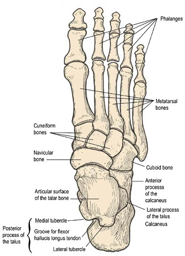
The navicular bone is located on the medial (inside) aspect of the foot between the talus and three cuneiform bones. It is an important bone that has many soft tissue attachments to it, most notably the muscle tibialis posterior (arch stabilising muscle) and the spring ligament (which also helps support the foot arch). Due to the poor blood supply to in this area, healing of this bone can be difficult and slow (Standring, 2015).

Clinical presentation
Often there is no one incident that will directly correlate to a navicular fracture. The individual may experience may be a gradual onset of a vague, dull ache in the mid-foot. It will begin to only effect the person while they are doing high load activities and dissipate with rest. As the condition progresses the dull ache will become more prominent even when not completing high load activities, such as walking or even when resting (Gross & Nunley, 2015). The major point of tenderness and pain is usually described as the ‘N-spot’ or navicular notch on the navicular.
When testing the foot and ankle clinically for a navicular stress response, there will usually be minimal to no loss of range of motion at the ankle joint although there may be pain with inversion and eversion. It can take up to 6 months for the niggling pain to become a problem that treatment is sort out for. Unfortunately, by the time that has come round it has progressed to a stress fracture rather than just bone oedema or a stress reaction (Patel, Christopher, Drakos, & O'Malley, 2020).
Injury to the navicular falls on the bone stress continuum meaning that an injury can be anywhere from bony oedema all the way though to complete fracture of the bone. Therefore, presentation and treatment will vary depending on the severity of the injury. If an injury to the navicular is suspected, it is recommended to image the foot to confirm the extent of injury in the navicular. The literature has shown that X-Rays are not very accurate at diagnosing a stress fracture (Harris & Harris, 2016). Currently in the literature, it has been found that both MRI and CT-scans can detect navicular stress fractures. CT-scans are unable to detect a stress reaction whereas an MRI can while also able to diagnosis other reasons for pain. Although a CT scan is said to be more useful when comparing fracture healing. Therefore, it is recommended to MRI the foot to get a clear understanding of the pathology (Harris & Harris, 2016).
Treatment
Management of a navicular bone injury is dependent on the severity of the injury. The most important factor is determining what led to the stress fracture and what changes can be made to ensure correct load management going forward. Treatment can be either operative or non-operative, which can be determined by the stability of the mid-foot. A metanalysis by Torg 2010 compared the outcomes of patients that had surgery compared to patients that had non-operative treatment. Non-operative treatment was broken up into either a non-weight baring cast (NWB) or in a weight baring cast (WB). All patients all had either a partial or non-displaced complete fracture.
The results found that 96% of patients treated with NWB cast for 6 weeks had a successful return to activity with an average of 4.9 months. Compared to 77% of patients (although these papers had a much lower total number of patients) who had less than 6 weeks of a NWB cast successfully returned to activity although the time was reduced to 3.7 months. When comparing to a weight bearing cast only 47% of patients had a successful outcome and return to activity and the time was on average 5.7 months. When compared to surgical intervention 82% had a successful return to activity in an average of 5.2 months (Torg, Moyer, Gaughan, & Boden, 2010).
Therefore, it is recommended that treatment consists of at least 6 weeks in a NWB boot to allow the navicular to heal. Reimaging of the bone can also be completed to confirm healing to decrease the risk of refracture.
Please seek medical advice from your Chiropractor, Physiotherapist or Sports doctor if symptoms do not resolve or become severe. Remember, the quicker you take care of your pain, the quicker you will be back to doing what you love.
References:
Gross, C. E., & Nunley, J. A. (2015). Navicular stress fractures. Foot & ankle international, 36(9), 1117-1122.
Harris, G., & Harris, C. (2016). Imaging of tarsal navicular stress injury with a focus on MRI: A pictorial essay. Journal of medical imaging and radiation oncology, 60(3), 359-364.
Mallee, W. H., Weel, H., van Dijk, C. N., van Tulder, M. W., Kerkhoffs, G. M., & Lin, C.-W. C. (2015). Surgical versus conservative treatment for high-risk stress fractures of the lower leg (anterior tibial cortex, navicular and fifth metatarsal base): a systematic review. British Journal of Sports Medicine, 49(6), 370-376.
Patel, K. A., Christopher, Z. K., Drakos, M. C., & O'Malley, M. J. (2020). Navicular Stress Fractures. JAAOS-Journal of the American Academy of Orthopaedic Surgeons, 10.5435.
Snyder, R. A., Koester, M. C., & Dunn, W. R. (2006). Epidemiology of stress fractures. Clinics in Sports Medicine, 25(1), 37-52.
Standring, S. (2015). Gray's anatomy e-book: the anatomical basis of clinical practice: Elsevier Health Sciences.
Torg, J. S., Moyer, J., Gaughan, J. P., & Boden, B. P. (2010). Management of tarsal navicular stress fractures: conservative versus surgical treatment: a meta-analysis. The American journal of sports medicine, 38(5), 1048-1053.
